A team of physicians in New York recently made a breakthrough in the diagnosis and treatment of a challenging cardiology patient. Excelsior Medical stated that the patient had a “complicated and serious medical condition” and was unable to receive a definitive diagnosis at a major New York medical center before getting discharged.
Excelsior Integrated Medical Group (Excelsior Medical) is a multi-specialty medical group based in New York dedicated to providing “high-quality care, value-based medical care delivery, population management, [and] healthcare cost reduction.” The group consists of over 70 providers spread across 19 divisions, 21 primary care sites, and 27 specialty sites.
The patient, Mr. AM, initially met with primary care specialist Dr. Tina Wong for hospital follow-up to discuss his diagnoses of non-ST elevation myocardial infarction (NSTEMI) and congestive heart failure. At the hospital, he received a diagnostic catheterization showing “normal coronary arteries” and “decreased overall left ventricular function of about 40%.” In other words, his heart vessels were not occluded, but his heart function was lower than normal due to an unknown etiology.
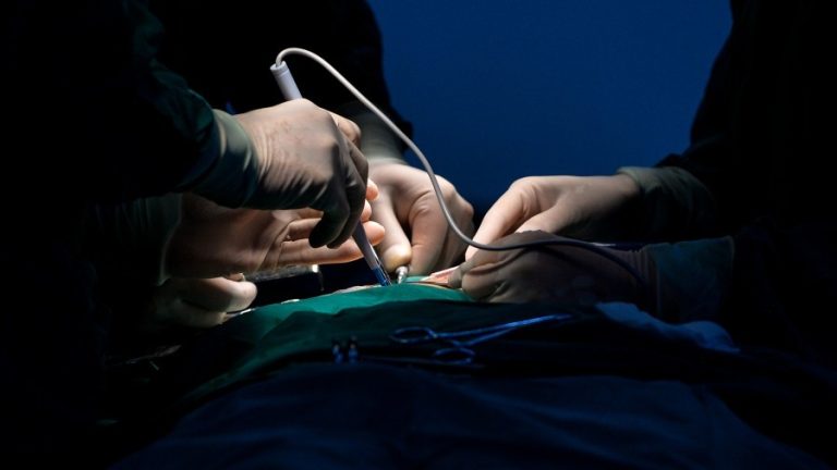
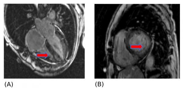
The patient was promptly referred to cardiologist Dr. Michael Poon, who evaluated the echocardiogram study. Given Mr. AM’s “compromised left ventricular function,” with no obvious abnormalities of the heart wall or valves, Poon decided to order a cardiac magnetic resonance imaging (MRI) scan. Poon is the regional director of Northwell Health Advanced Cardiac Imaging and the MRI was performed at Lenox Hill Greenwich Village Advanced Cardiac Imaging Center.
The findings from the MRI study were invaluable for guiding Poon’s next steps. The images “showed the classic pattern of cardiac amyloidosis with global subendocardial late enhancement.” According to Johns Hopkins, the condition “occurs when plaques of a protein byproduct called amyloid build up in heart muscle, affecting its ability to pump blood.”
Success
You are now signed up for our newsletter
Success
Check your email to complete sign up
The patient was immediately referred to the group’s hematology expert, Dr. Songchuan Guo, for further evaluation of the presumptive diagnosis of systemic amyloidosis. Hematologists specialize in blood as it relates to health and disease. Mr. AM was found to have mild anemia or a decreased proportion of red blood cells. In addition, a Serum Protein Electrophoresis (SPEP) study revealed a monoclonal (M) spike of IgA Lambda (1.8 g/dl), which was confirmed by high levels of IgA antibodies (3162 mg/dl) and Lambda light chains (224 mg/dl), pieces of antibodies made by white blood cells, in Mr. AM’s blood. No significant abnormalities in kidney and liver function were found.
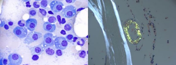
Subsequently, Guo ordered a bone marrow biopsy, in which a sample of the patient’s bone was taken for analysis. Cancer with an abnormally high proportion of plasma cells, a type of white blood cell, was confirmed. Congo Red staining of the sample revealed “deposits within vascular walls, consistent with amyloid deposition.”
Within one week of consulting Guo, Mr. AM was diagnosed with systemic amyloidosis based on clinical presentation and laboratory and pathology findings. The next day, he was started on bortezomib-based chemotherapy, and “tolerated the treatment very well.” Furthermore, he was referred to a transplant specialist in hopes of becoming a “future stem cell transplantation recipient.”
Mr. AM expressed his sincere gratitude to the Excelsior Integrated Medical Group, which exhibited efficient coordination between physicians in primary care and multiple specialty divisions. With the lack of a definitive diagnosis during his hospital stay, the patient was pleased to receive clarity about his condition and begin a treatment plan.
Distinguished cardiologist
Dr. Michael Poon, who was instrumental in diagnosing and treating Mr. AM, is a prestigious US-trained cardiologist who immigrated to New York City from Hong Kong at the age of 15. He received his Medical Doctor (MD) degree from Mount Sinai School of Medicine and was at the top of his class as part of the Alpha Omega Alpha (AOA) Honor Medical Society.

During his internal medicine and cardiology fellow training at Mount Sinai Hospital, he won several prestigious awards, including Young Investigator Awards from the American Heart Association (AHA) and American College of Cardiology (ACC), and the AHA Clinician Scientist Award.
Before joining Excelsior Integrated Medical Group, Dr. Poon was System Director of Research, Cardiac Imaging, and Clinical transformation of Northwell Health and Director of Noninvasive Cardiac Imaging at Lenox Hill Hospital and holds professorship titles in five academic departments.
Poon’s clinical and scientific accomplishments include the discovery of the “antiproliferative property of Rapamycin in 1994” and groundbreaking research in the area of drug-eluting coronary stent treatment for obstructive coronary artery disease (CAD). He “started the field of coronary computed tomographic angiography (CCTA)” in America and spearheaded the use of cardiac MRI for the evaluation of right-sided heart failure and pulmonary hypertension.
Furthermore, he led efforts to use CCTA in the Emergency Room triage of acute chest pain at Stony Brook University Medical Center and Fractional Flow Reserve CT (FFRct) as an everyday clinical tool at Northwell Health. Most recently, he “pioneered the use of CCTA” during the coronavirus pandemic and “led the national movement to start using CCTA as the preferred initial diagnostic test for the evaluation of stable chest pain.”
Dr. Michael Poon practices in New York at the following clinics:
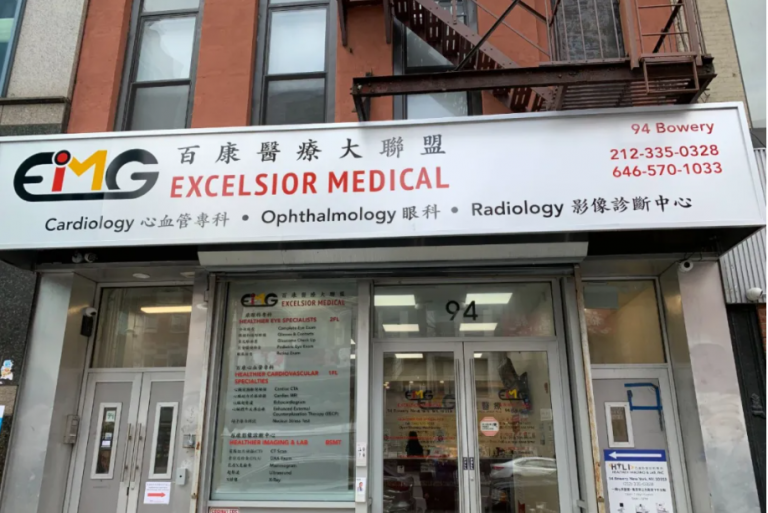
94 Bowery
New York, NY 10013
Tel:(212) 925-4088
758 61st St
Brooklyn, NY 11220
Tel:(212)925-4088
38-08 Union St #13A
Flushing, NY 11354
Tel:(212) 925-4088